|
|
|
Structural biology of plant macromolecules III
|
|
|
| Crystal structure of cellulose Iƒ¿ and IƒÀ |
|
|
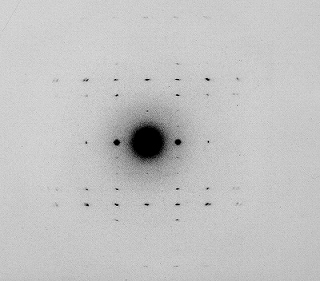
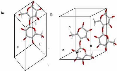
First X-ray Laue diagram was taken by Nishikawa and Ono in 1913. Since then the concepts that all the cellulose samples should be characterized by a uniform cellulose I model. This concept has been revised when high resolution solid state NMR became available and a range of cellulose samples were characterized. Finally, electron micro- diffraction study confirmed that cellulose I is a mixture of cellulose Iƒ¿ and IƒÀ, and concluded that the former adopts tricilinic one chain unit cell while the latter adopts
monoclinic two chain unit cell, respectively.
|

|
|

|
Sugiyama J, Vuong R, Chanzy H: An electron diffraction study on the two crystalline phases occurring in native cellulose from algal cell wall. Macromolecules, 24, 4168-4175, 1991
Sugiyama J, Persson J, Chanzy H: A combined infrared and electron diffraction study of the polymorphism of native celluloses. Macromolecules, 24, 2461-2466, 1991
Sugiyama J, Okano T, Yamamoto H, Horii F: Transformation of Valonia cellulose crystals by an alkaline hydrothermal treatment. Macromolecules, 23, 3196-3198, 1990
|
|
|
H-bonding networks are now established by state of arts SR X-ray and neutron fiber diffraction studies.
Wada M, Heux L, Sugiyama J: Polymorphism of cellulose I family: reinvestigation of cellulose IVI. Biomacromolecules, 5, 1385-1391, 2004
Nishiyama Y, Sugiyama J, Chanzy H, Langan P: The crystal structure and hydrogen bonding system in cellulose Iƒ¿ from synchrotron X-ray and neutron fiber diffraction. J Am Chem Soc, 125, 14300-14306, 2003
Imai T, Sugiyama J, Itoh T, Horii F: Almost pure Ia cellulose in the cell wall of Glaucocystis. J Struct Biol, 127, 248-257, 1999
|
|
| |